Objective: design and construction of an automated high-resolution device for automatic 3D modeling and analysis of the patient's skin surface and for the identification of pigmented lesions for secondary prevention of skin cancer.
The aim of the project is to develop a high-resolution device for automatic 3D modelling and analysis of the patient's skin surface to identify pigmented lesions (so-called moles) for the secondary prevention of skin cancer (in particular melanoma).
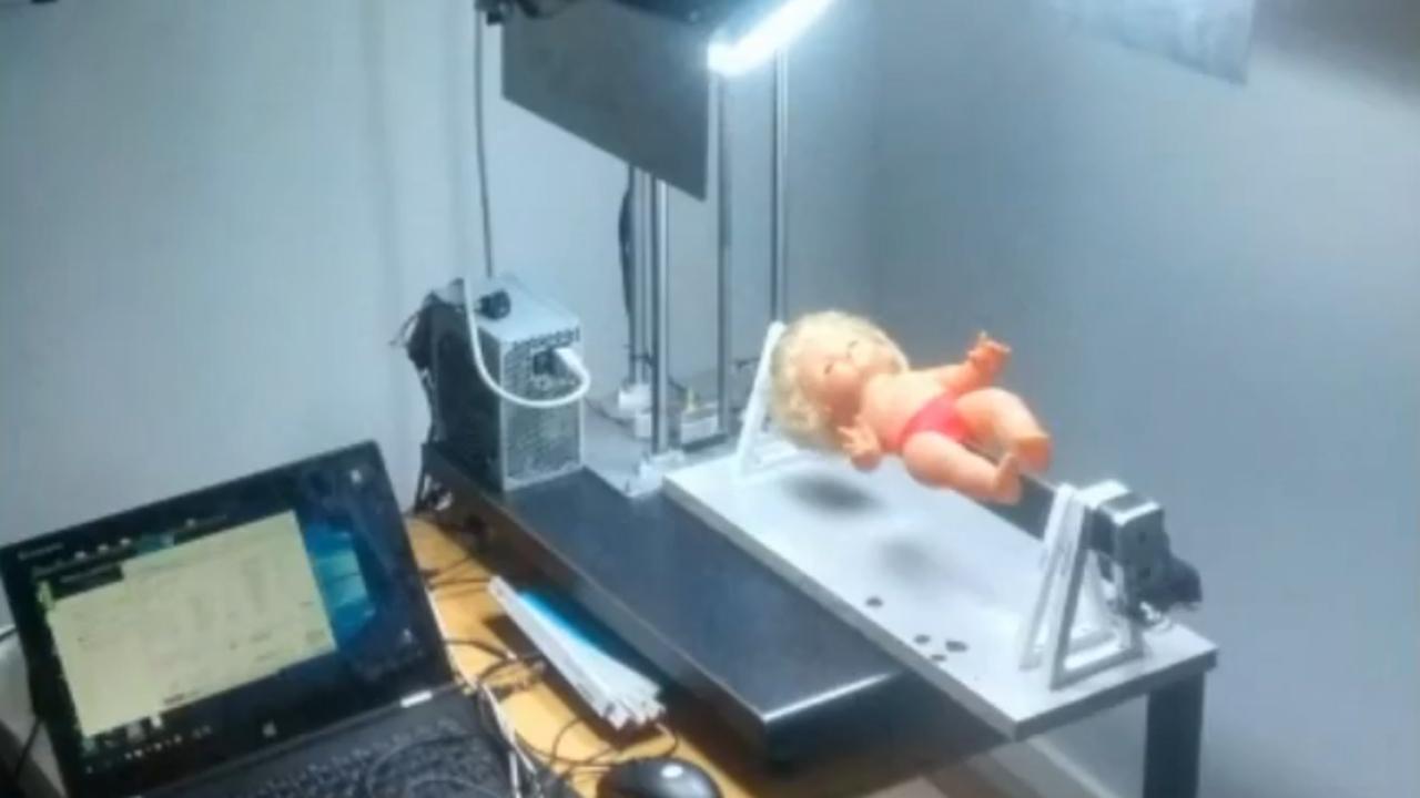
The device will be a platform to which a boom with camera(s) (1 to 6) will be attached. The camera (or several cameras) will move in a linear (up and down) and angular (around the patient) motion, taking detailed images of the body (at least several dozen shots), from which an accurate 3D map of the body will be created. Each image will depict a section of skin with a small area (e.g. 30x20cm, approximately 290 pixels for every 1 mm2 of skin). By increasing the precision of the imaged details (higher imaging resolution, measurement of image depth using structured light), it will be possible to develop and apply new algorithms for nevus detection, e.g. related to nevus edge search, level set method, field strength gradient method, etc.
New nevus matching algorithms will be developed, which will use information about the position of the nevus in the 3D model, the distance between the matched nevus and neighbouring nevi, as well as its parameters such as shape, size. Advanced algorithms will be developed to reduce the influence of factors such as lighting irregularities, changes in the patient's skin tone, hairiness, etc. Measurements using polarised light and measurements using colour filters will be carried out to increase the efficiency of extracting information from images.
As a result, the new device will allow the detection of new nevi and the analysis of the alteration of existing nevi, compared to competing solutions:
- with significantly higher efficiency
- in a shorter time
- at a lower price.
The project is being implemented within a group of entities.
Project leader: Mariola Janczy, Skopia Estetic Clinic Sp. z o.o., KRAKÓW
Head of research team at the PŁ: prof. dr hab. Michał Strzelecki
Years: 2018 – 2022
Projekt pt. "Opracowanie urządzenia do wspomagania wczesnej diagnostyki znamion skórnych, w tym czerniaka, za pomocą metod wizji komputerowej, modelowania przestrzennego, analizy porównawczej i klasyfikacji", realizowany zgodnie z umową o dofinasowanie nr POIR.04.01.04-00-0125/18-00 z dnia 27.12.2018 r.
The project is co-financed by the European Union - European Regional Development Fund under the Operational Programme Intelligent Development.
Value of the project for the Lodz University of Technology - 1.272,200 zł
Contribution from the European Funds - 1.272.200,00 zł
The results of the project to date have been presented in publications:
- Strzelecki, M.H.; Strąkowska, M.; Kozłowski, M.; Urbańczyk, T.; Wielowieyska-Szybińska, D.; Kociołek, M. Skin Lesion Detection Algorithms in Whole Body Images. Sensors 2021, 21, 6639.
- Szczypiński, P.M.; Sprawka, K. Orthorectification of Skin Nevi Images by Means of 3D Model of the Human Body. Sensors 2021, 21, 8367.