Can the analysis of X-ray images of adjacent bones help detect hip joint arthrosis? 🤔
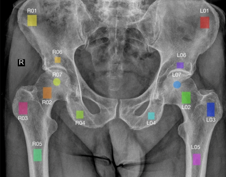
👉 Prof. Michał Strzelecki from our Institute, together with 👉 Prof. Rafał Obuchowicz from Jagiellonian University Collegium Medicum and 👉 Sebastian Borukało, a student at Lodz University of Technology, developed a deep learning model capable of automatically assessing the presence of degenerative changes in the hip joint based on X-ray images.
The study shows that even fragments of the pelvic and femoral bones contain valuable information about the condition of the hip joint - and that applying proper image preprocessing (e.g., CLAHE, Z-score normalization) significantly improves detection performance.
🔬 Article: “Assessment of hip joint arthrosis based on the X-ray images of adjacent bones”
📈 Results: balanced accuracy, sensitivity, and specificity ≈ 0.7
💡 Conclusion: structural changes in the bones are closely related to hip joint arthrosis - and AI may help detect them earlier than ever before.
✒️ We encourage you to read the full paper:
S. Borukało, M. Strzelecki and R. Obuchowicz, "Assessment of hip joint arthrosis based on the X-ray images of adjacent bones," 2025 Signal Processing: Algorithms, Architectures, Arrangements, and Applications (SPA), Poznan, Poland, 2025, pp. 159-164, doi: 10.23919/SPA65537.2025.11215118.