Numerical modeling of the cerebral arterial and venous blood-vessel system in macro- and mesoscale based on 3D MRI data
The aim of the project is to investigate the impact of basic phenomena used in magnetic resonance equipment (MRA) on the accuracy of modelling and parameterisation of the cerebral vascular system.
Two hypotheses are planned to be proven.
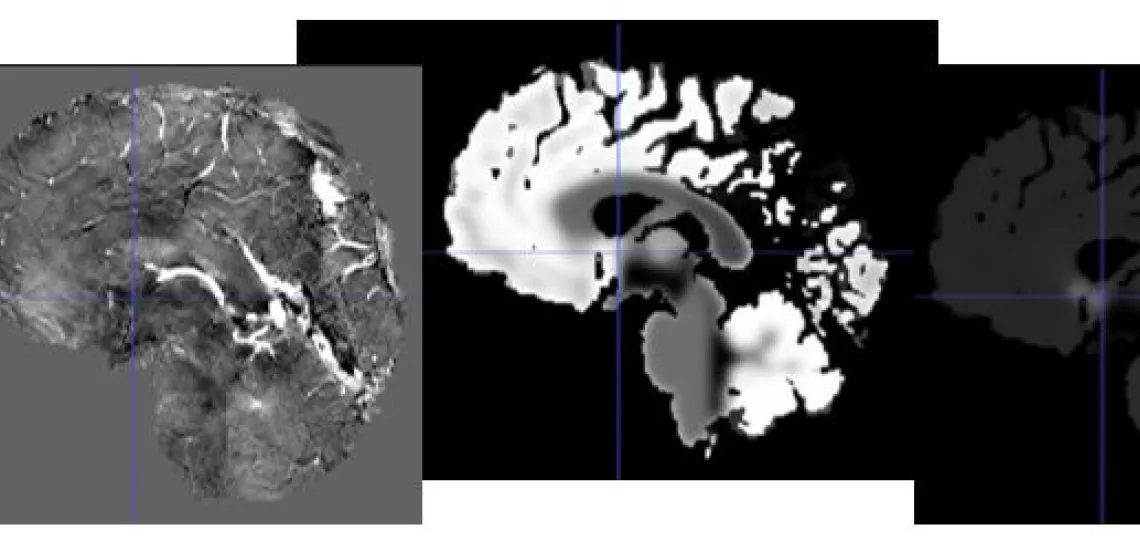
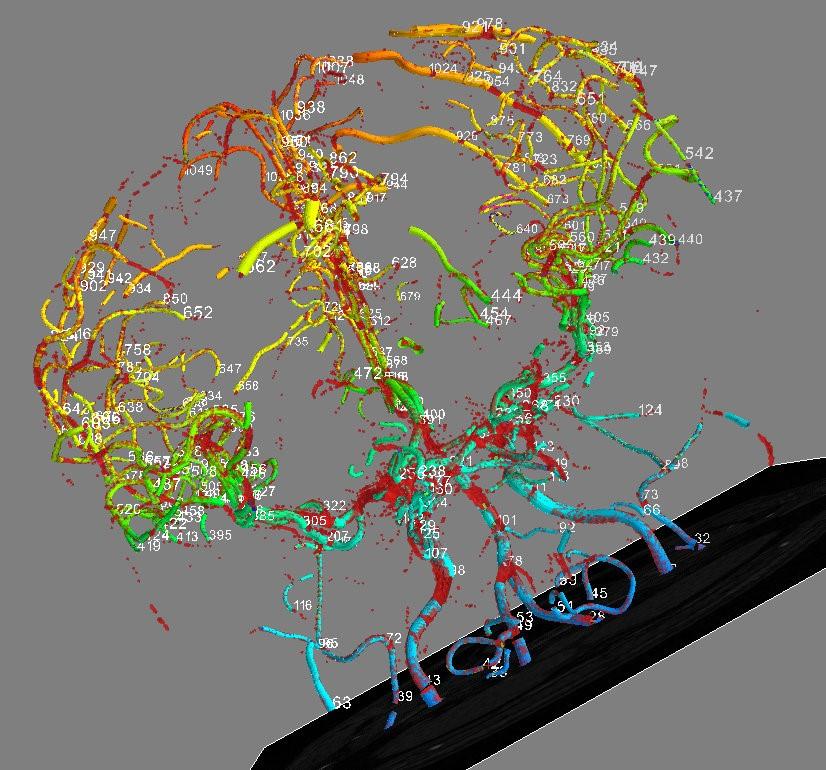
As the spatial resolution of RM images for a fixed magnetic field strength is limited, geometric models of the vessels can only be built for large diameter branches (relative to the side length of the image voxel). It is also known that thinner vessels are a continuous extension of thick vessels, and that areas filled with them can be quantitatively characterised using statistical texture parameters or SWI-measured vessel density maps. These observations allow us to formulate the hypothesis that attaching numerically synthesised thin vessel trees (with statistical parameters matched to images) to the branches of a geometric model of large-diameter vessels will allow continuous modelling of vessel tree properties over a much wider diameter scale than is currently possible.
The second hypothesis is that the combination of various 3D image segmentation methods (including the level method with mathematical morphology and regularisation of the relevant equations) will allow models to be built that are robust to the artefacts (including contrast spikes) that accompany sequences used to image arteries and veins.
Project Leader and Researchers: prof. A. Materka (project leader), prof. J. R. Reichenbach, dr A. Deistung, dr hab. M. H. Strzelecki, dr hab. P. M. Szczypiński, dr hab. A. J. Majos, dr inż. M. Kociński, dr inż. A. J. Klepaczko, mgr A. Chmielewska-Pawlicka.
Years: 2013 – 2016
Project type: NCN-MN, NCN HARMONIA Grant; fundamental research
Key publications resulting from the project:
- A. Klepaczko, P. Szczypiński, G. Dwojakowski, M. Strzelecki, A. Materka, Computer Simulation of Magnetic Resonance Angiography Imaging: Model Description and Validation, PLoS One 2014;9(4):e93689, DOI: 10.1371/journal.pone.0093689 (IF=3,534).
- A. Klepaczko, A. Materka, P. Szczypiński, M. Strzelecki, Numerical Modeling of MR Angiography for Quantitative Validation of Image-Driven Assessment of Carotid Stenosis, IEEE Transactions on Nuclear Science, 2015;62(3):619-627, DOI: 10.1109/TNS.2015.2433925 (IF=1,283).
- M. Kociński, J. Blumenfeld, A. Materka, A. Deistung, B. Serres, JR. Reichenbach (2015) Automated centerline-based modeling of tubular blood vessel segments from 3D MRA. Magnetic Resonance Materials in Physics, Biology and Medicine MAGMA, Vol 28, Suppl 1, Oct 2015, 32nd Annual Scientific Meeting, Edinburgh, October 1-3 2015, pp. S381-S382, DOI: 10.1007/s10334-015-0489-0, © Springer.
- J. Blumenfeld, M. Kociński, A. Materka (2015) A centerline-based algorithm for estimation of blood vessels radii from 3D raster images. IEEE SPA 2015 (Int. Conf on Signal Processing, Algorithms, Arrangements and Applications), 26-28 Sept 2015, Poznań, pp. 38-43, © IEEE.
- A. Materka, M. Kocinski, J. Blumenfeld, A. Klepaczko, A. Deistung, B. Serres, JR. Reichenbach (2015) Automated Modeling of Tubular Blood Vessels in 3D MR Angiography Images, 9th International Symposium on Image and Signal Processing and Analysis (ISPA 2015) September 7-9, 2015, Zagreb, Croatia, pp. 56-61, © IEEE.
Image